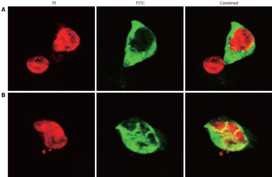
Figure 1.
SP-TAT-apoptin expression in HepG2 cells (× 1000). Cells transfected with plenti6/V5-D-TOPO/SP-TAT-apoptin plasmid and fixed at 24 h (A) and 48 h (B) post-transfection. Recombinant apoptin detected by anti-V5-FITC antibody is shown in green and cell nuclei stained by PI in red. Apoptin protein showed a diffuse pattern in the cytoplasm at 24 h post-transfection, and in the nucleus at 48 h.

