Figure 3.
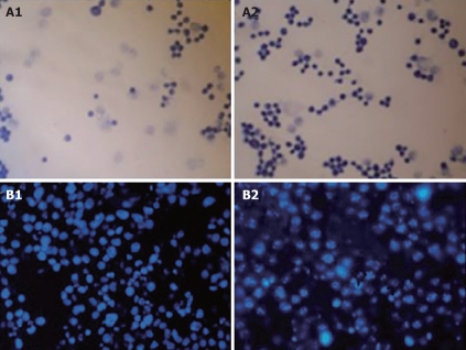
Cytotoxicity of SP-TAT-apoptin compared to SP-TAT. A: Micrographs of HepG2 cells transfected with SP-TAT-apoptin construct stained by Apoptotic/Necrotic Cell Detection Kit. An inverted microscope (× 400) was used. The nuclei of apoptotic cells were stained deep blue. A1 and A2: HepG2 cells at 24 and 72 h post-transfection; B: HepG2 cells stained with DAPI and observed by fluorescence microscopy (× 400). B1: HepG2 cells 72 h after transfection with plenti6/V5-D-TOPO/SP-TAT-GFP plasmid; B2: HepG2 cells 72 h after transfection with plenti6/V5-D-TOPO/SP-TAT-apoptin. Arrow indicates apoptotic cells.

