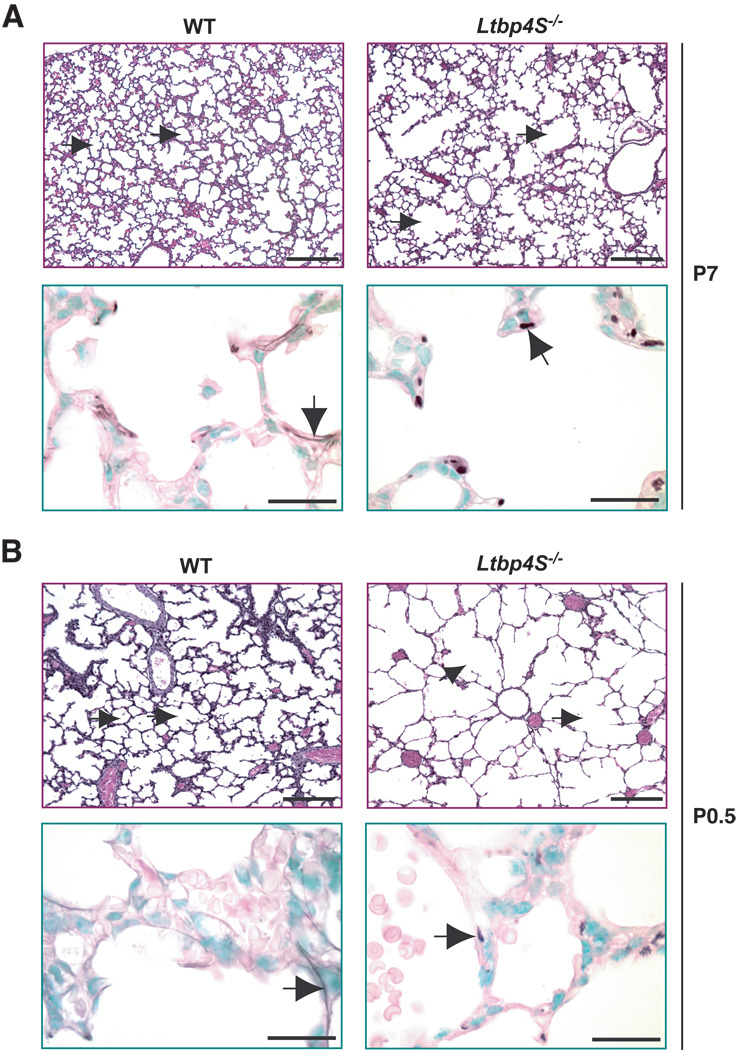
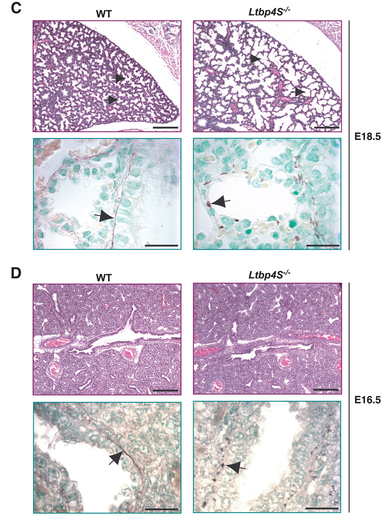
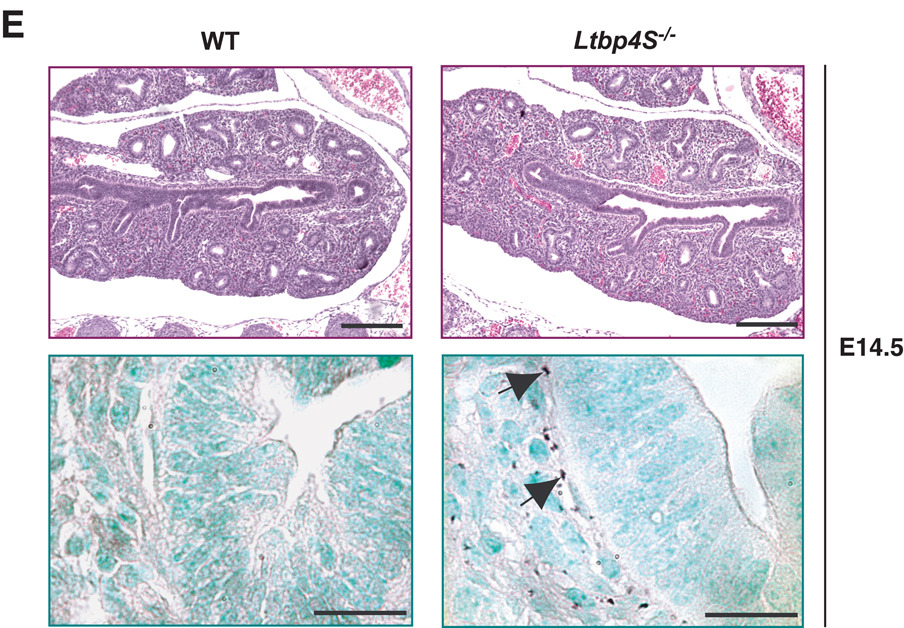
Figure 1.
Defective terminal air-sac septation and elastic fiber formation in Ltbp4S−/− lungs. Upper panels show lung sections stained with H&E, lower panels present orcinol-new fucsin staining of elastin. The arrows in upper panels point to terminal air-sacs; the arrows in the lower panels point to elastin. A. P7 In WT lungs at P7 terminal air-sacs are divided into small units by the process of alveolarization, whereas in Ltbp4S−/− lungs alveolarization is not uniform yielding regions with large terminal air-sacs. Elastin in the WT alveolar walls appears fibrillar, whereas in the Ltbp4S−/− alveolar walls only globules of elastin were observed. B. P0.5 At P0.5, the terminal air-sacs in Ltbp4S−/− lungs are much larger than in WT lungs. The differences in elastin organization between WT and mutant lungs are similar to those illustrated in A. C. E18.5 The difference in terminal air-sac septation and in elastin organization between WT and Ltbp4S−/− lungs was already obvious at E18.5. D and E. E16.5 and E14.5 No differences in WT and Ltbp4S−/− lung histology were observed both at E16.5 and E14.5. However the differences in elastin organization were observed at E16.5 (D) and E14.5 (E). Bars: upper panels − 200 µm, lower panels − 20 µm.


