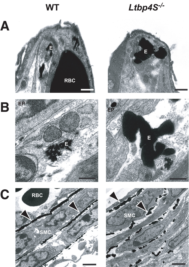
Figure 2.
Electron micrography of P0.5 WT and Ltbp4S−/− lungs. A Alveolar tips. B Airways. C Blood vessels. The lack of continuous lamellae in the mutant blood vessels is clear in C where there are essentially no ordered lamellae. E – elastin, RBC – red blood cell, SMC - smooth muscle cell, EP – epithelial cell. Arrows point to elastic lamellae in the walls of blood vessels. Bars: A −0.5 m, B - 0.2 µm, C - 2 µm.

