Figure 4.
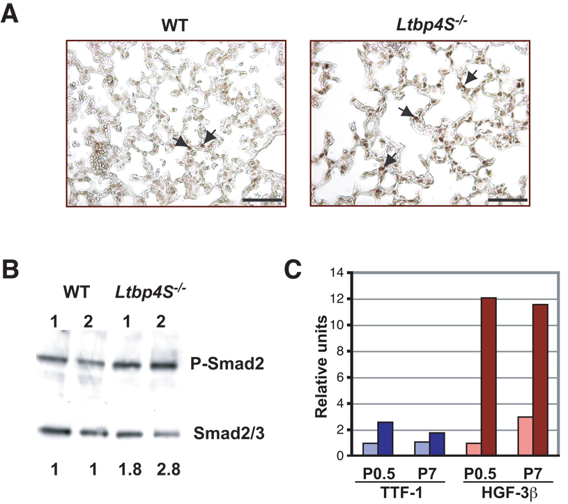
Increased TGF-β signaling in Ltbp4S−/− lungs. A Immunohistochemistry with P-Smad2 antibody revealed a higher number of positive cell nuclei in Ltbp4S−/− lungs. The arrows point to the P-Smad2 positive nuclei. B Quantitative Western Blot analysis of P-Smad2 in the lungs from 2 WT and 2 Ltbp4S−/− P7 mice. The numbers at the bottom indicate the ratio of the intensity of P-Smad2 vs. Smad2/3 in the Ltbp4S−/− samples normalized to the P-Smad2 to Smad2/3 ratio in the WT samples. The ratio of the intensity of the P-Smad2 and the Smad2/3 bands was equivalent in both WT samples. The result shown is representative of four experiments using different animals. C Graphic representation of expression of TTF and HNF-3h in WT and mutant lungs. The expression of both genes was enhanced in Ltbp4S−/− mice indicating increased TGF-β levels. Bar: 5 µm.

