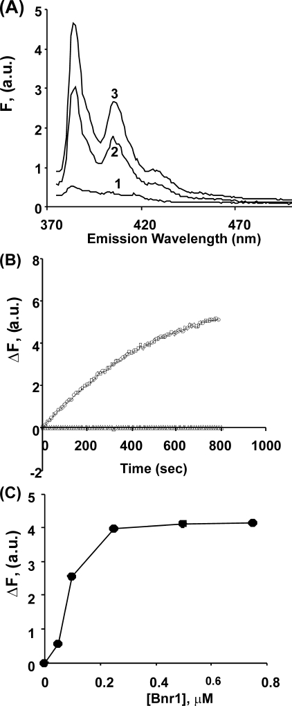
FIGURE 1.
Interaction of Bnr1 with pyrene-labeled Mg-G-actin. Panel A, aliquots of 0.5 μm pyrene-labeled Mg-G-actin were combined with increasing amounts of Bnr1 (line 1: 0 μm, line 2: 0.1 μm, and line 3: 0.5 μm) in Mg-G-buffer conditions (10 mm Tris-HCl pH 7.5, 0.2 mm MgCl2, 0.2 mm ATP, 100 μm EGTA, and 1 mm DTT), and the solutions were incubated at room temperature for 15 min at room temperature. The fluorescence emission spectra were recorded from 375 to 500 nm following excitation at 365 nm. Panel B, pyrene-labeled Mg G-actin was combined with Bnr1 (Δ: no Bnr1, and ○: 0.5 μm) in Mg-G-buffer, and the increase in pyrene fluorescence intensity at emission wavelength 385 nm was recorded over time. Excitation wavelength was 365 nm. Panel C, net change pyrene fluorescence intensity at emission wavelength 385 nm from panel A was plotted against the concentration of Bnr1 as described under “Experimental Procedures.” The experiments shown in each panel have been repeated twice with essentially the same results.

