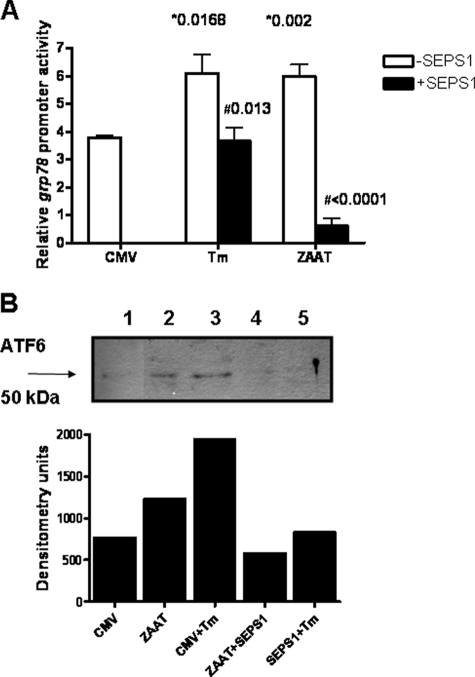
FIGURE 2.
SEPS1 inhibits ZAAT-induced activation of UPR. A, triplicate samples of HepG2 cells (3 × 10⁵) were cotransfected with an empty vector (pCMV) or pSEPSI; pZAAT; an inducible grp78 promoter-linked (firefly) luciferase reporter plasmid; and pRLSV40. Cells were treated with tunicamycin (Tm; 10 μg/ml) or DMSO for 16 h. Lysates were prepared, and luciferase production from both plasmids was quantified by luminometry using specific substrates. Relative grp78 promoter activity is shown (*, versus CMV; #, versus minus SEPS1). B, HepG2 cells (3 × 10⁵) were transfected with pCMV or pZAAT, treated with tunicamycin (Tm; 10 μg/ml) or DMSO. Western immunoblotting for ATF6 was carried out on cell lysates, and densitometry of the resulting blot is also shown. Lane 1, empty vector (CMV); lane 2, pZAAT; lane 3, CMV + tunicamycin; lane 4, pZAAT + pSEPS1; lane 5, CMV + tunicamycin + pSEPS1. Data shown are representative of three experiments.

