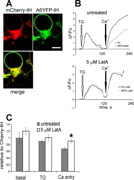
FIGURE 10.
Destabilization of actin cytoskeleton with latrunculin A abolishes the annexin A6YFP-tH-mediated decrease of Ca2+ entry. A, HEK293 cells co-expressing annexin A6YFP-tH and mCherry-tH were treated with 5 μm Lat A for 2 h, and examined in the confocal microscope. Bar = 5 μm. B, co-cultured HEK293 cells expressing annexin A6GFP-tH (A6YH) or mCherry-tH (CH) were loaded with Fluo-3/AM, and responses to the application of 1 μm TG followed by stimulation of SOCE recorded by calcium imaging. Representative recordings from untreated cells (top), and from cells incubated in 5 μm Lat A for 1 h before SOCE activation (bottom) are shown. C, basal [Ca2+]i levels, TG-stimulated ER release and Ca2+ entry in A6YFP-tH cells treated with 5 μm Lat A for 1 h, or in untreated A6YFP-tH cells were expressed relative to the responses of the control, mCherry-tH cells. The graph shows an average of three independent experiments (n = 10 arbitrary fields each measured for untreated and Lat A-treated coverslips) ± S.E. The difference in relative SOCE levels between untreated and Lat A-treated A6YFP-tH cells was statistically significant (*, p < 0.05).

