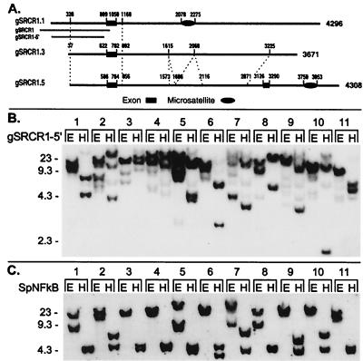
Figure 3.
Genomic diversity in type 1 SRCR genes. (A) Schematic presentation of similarity among three genomic clones that were detected with an SRCR type 1 5′ flanking probe. Domains displaying high similarity among the sequences are connected by dashed lines. Exon 1 corresponds to the 5′ UTR and leader peptide in SpSRCR1 (nucleotides 1–161), and exon 2 in gSRCR1.5 corresponds to the N-terminal half of the SRCR1 domain (nucleotides 1,939–2,103 in SpSRCR1). Composite dinucleotide CT microsatellites are denoted by an oval symbol. Corresponding regions of the SRCR1 5′ flanking probes are marked underneath gSRCR1.1: gSRCR1 (1,078-bp, 46-nt overlap with the first exon) and gSRCR1–5′ (805 bp, nucleotides 147–952 in gSRCR1). (B) Genome blot with the gSRCR1–5′ probe. (C) Genome blot probed with the single copy gene marker SpNFkB. The SpNFkB probe corresponds to a region located at the center of the Rel homology domain. Blot is the same as that shown in Fig. 2 (E; EcoRI; H; HindIII). Numbers to the left of the blots indicate length (kb).

