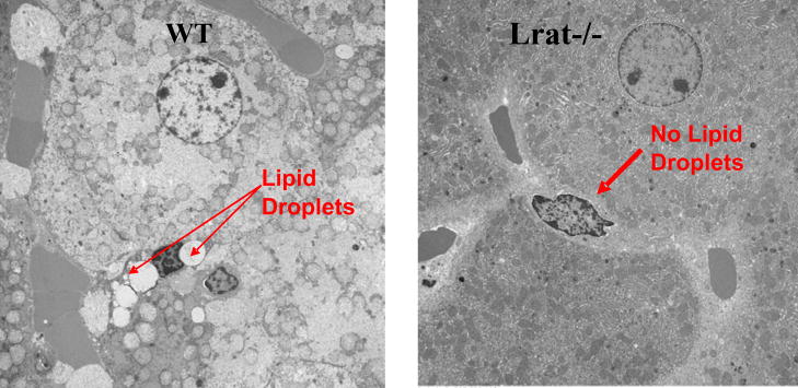
Fig. 4.
The hepatic stellate cells of Lrat−/− mice lack lipid droplets that are a morphologic hallmark of these cells. Liver sections were prepared from 3-month-old male wild type (WT) and Lrat−/− mice. The electron micrographs show the presence of characteristic retinyl ester-containing lipid droplets in hepatic stellate cells in wild type mice (left panel) and their absence in livers from Lrat−/− mice (right panel). The arrows indicate the presence (WT) and absence (Lrat−/−) of lipid droplets in hepatic stellate cells. The large adjoining cells are hepatocytes. Taken from [32].

