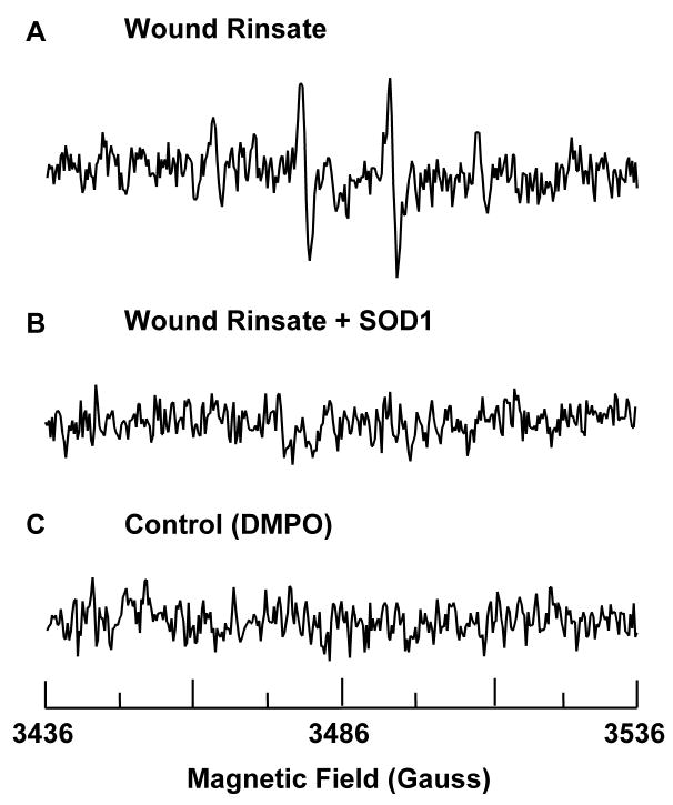
Figure 2.
Presence of superoxide at wound site. (A) The spectra shows presence of spin adduct DMPO-OH in wound rinsate. 50 μl of 1 M DMPO was topically applied to 2 days old 8 mm punch biopsy wounds. After 10 minutes wound rinsate was collected from the wound cavity, diluted 20X in PBS containing DTPA, and its EPR spectrum was recorded. (B) 5 μl of SOD1 (300 μM) was topically applied to the wound 10 minutes before application of DMPO and subsequent collection of wound rinsate. Addition of SOD1 quenches the EPR signal indicating that the source of signal is from superoxide. (C) Spectrum of DMPO in PBS. X-band EPR measurements were carried out using a quartz flat cell at room temperature. EPR instrument parameters used were as follows: microwave frequency 9.77 GHz; modulation frequency 100 kHz; modulation amplitude 1 G; microwave power 20 mW; number of scans 30; scan time 30 s; and time constant 81 ms.

