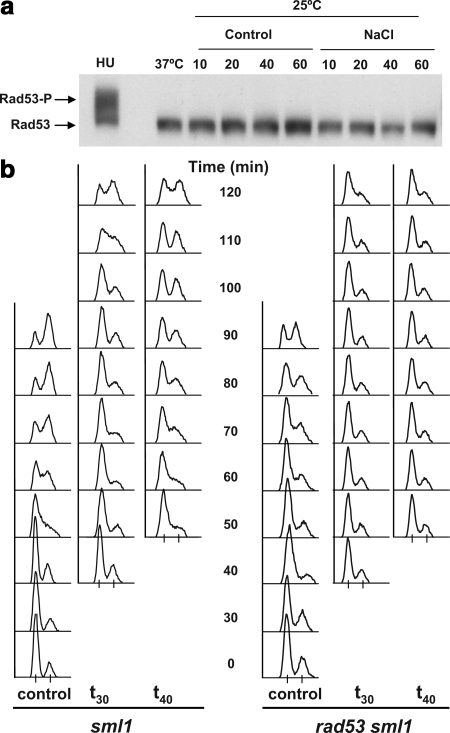
Figure 3.
The Rad53 DNA damage checkpoint is not involved in the Hog1 induced S phase delay. (a) The effector kinase Rad53 is not hyper-phosphorylated in response to osmostress. Exponentially growing cells with endogenously HA-tagged Rad53 were synchronized with α-fac for 3 h. Cells were washed free of the α-factor and stressed with 0.4 M NaCl either 20 min (left, t20) or 40 min (right, t40) after α-factor release. Samples were taken immediately before NaCl addition (0) and 10, 20, 40, and 60 min after it. Protein was extracted, separated on 7% gel, and Western blot analysis was performed with α-HA antibodies. Part of the culture was incubated with HU for 1 h immediately after release from α-factor (HU). Nonphosphorylated Rad53 is indicated by the bottom arrow and slower migrating phosphorylated Rad53 forms are present above in the HU sample (P-Rad53-HA). (b) Osmostressed rad53 cells delay in S phase. sml1 (left-hand side) and sml1 rad53 cells (right-hand side) were grown, synchronized with α-factor, and released as described in a. Cultures were subsequently osmostressed with 0.4 M NaCl 30 min (t30) or 40 min (t40) subsequent to α-factor release. Samples were collected and analyzed by FACS. The progression of a nonstressed culture is shown (control). Experiments were performed at least twice, and representative results are shown.

