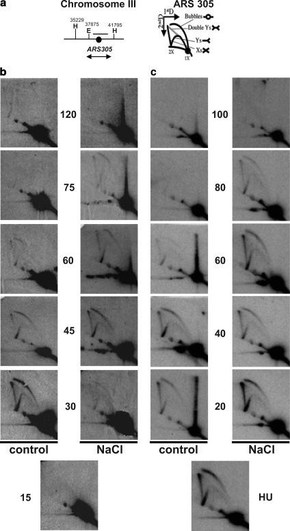
Figure 8.
Replication is altered in osmostressed cells. (a) Schemes of the chromosome III region around ARS305 analyzed (left) and of the migration pattern of the Y-, bubble-, double Y-, and X-shaped replication intermediates in 2D-gel electrophoresis (right). The region used as a probe in the hybridization experiments is indicated (b) Replication firing is delayed in cells osmostressed after α-fac synchronization. Exponentially growing sic1 bar1 cells growing at 25°C were synchronized in α-fac for 3 h and liberated into fresh medium (control). Half of the culture was exposed to 0.4 M NaCl 15 min after release from α-factor (NaCl). 2D-gel analysis of EcoRV–HindIII restriction fragment covering ARS305 is shown. (c) Cells osmostressed after HU synchronization take longer to replicate. Cells as described in a were synchronized for 1 h in 200 mM HU directly after washing away the α-fac. The culture was then liberated of HU into fresh medium (control) or medium supplemented with 0.4 M NaCl (NaCl). 2D-gel analysis was performed as described in a.

