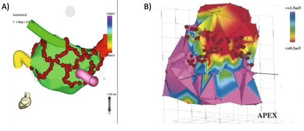
Figure 1.
Examples of electrospatial mapping guidance of complex arrhythmia ablation. A and B) Electrospatial surface maps generated by point-to-point contact mapping of the endocardial surface. The red circles are markers where ablation energy was delivered. A) Example of atrial fibrillation ablation in which ablations lesions are placed to encircle the pulmonary veins to prevent exit of arrhythmia triggering foci originating from the pulmonary veins. The pulmonary vein locations are marked by the colored "cartoon" tubes. B) Example of scar based ventricular tachycardia ablation in which linear lesions were places connecting scar (red) to normal tissue (purple) to interrupt the arrhythmia circuit. Figure 1A included with permission from The Journal of Cardiovascular Electrophysiology. (Calkins JCEP 2005; 13:53) Figure 1B included with permission from Circulation. (Marchlinski, Circ 2000; 101:1288).

