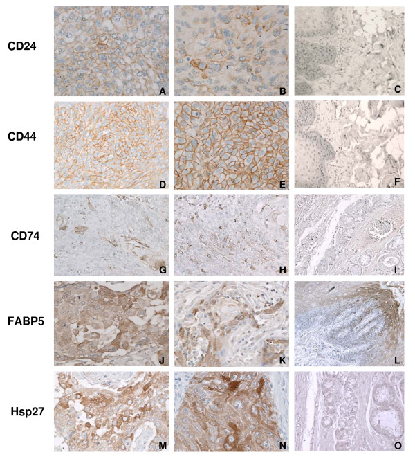
Figure 2.
Immunohistochemical analysis of CD24, CD44, CD74, FABP5, and Hsp27 in HNSCC samples. HNSCC tissue arrays contained 16 tumor samples from various locations and different stages. A and B are tumor samples and stained for CD24. D and E stained for CD44, and showed strong positive reactions in most tumor cells. Both CD24+ and CD44+ show cell surface staining, and CD24+ cells present in a small cluster of cells in the large tumor mass. (Magnification: ×200). G and H stained for CD74. Some of tumor cells showed CD74+. J and K stained for FABP5, and M and N stained for Hsp27. FABP5 shows strong cytoplasmic staining as well as Hsp27. C, F, I, L, O: tumor adjacent normal tissue. CD24, CD44, CD74, FABP5, and Hsp27 were negative in normal tissues, although FABP5 shows positive staining in the basal layer of dermis (Fig 2L), but negative in other areas; (Magnification: ×200). IgG was used as a negative control (Data not shown).

