Figure 2.
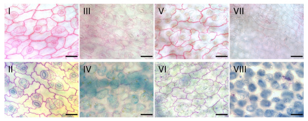
Degradation of cauliflower cell walls by extracellular produced pectic enzymes of seven R. solani AGs and one binucleate Rhizoctonia AG. Microscopic observations of pectic components in cauliflower cotyledones visualized with ruthenium red (I, III, V & VII) and toluidine blue (II, IV, VI & VIII) staining after 24 h incubation in sterile culture filtrate of liquid pectin medium inoculated with a sterile PDA plug as control treatment (I & II), inoculated with R. solani AG 3 (III & IV), inoculated with R. solani AG 4 HGII (V & VI), after 24 h incubation in sterile culture filtrate of liquid cauliflower medium inoculated with R. solani AG 4 HGII (VII & VIII). Scale bars = 50 μm.

