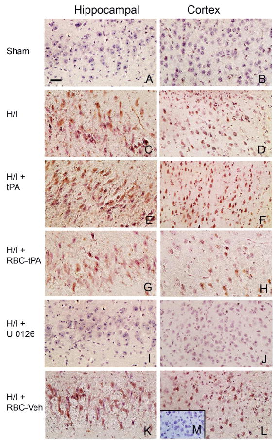
Figure 5.
tPA aggravates H/I induced ERK MAPK phosphorylation, while RBC-tPA blunts insult associated increases in ERK MAPK phosphorylation. Immunohistochemical data for phospho (activated) ERK MAPK obtained from piglets 4h after sham control (Panels A,B) or cerebral H/I and post-treatment 2 h after injury with either tPA (2 mg/kg), RBC-tPA (0.1 mg/kg), RBC-vehicle, or U 0126 (1 mg/kg). Abundant phospho ERK MAPK (4–5 on a 5 point scale) was observed in the hippocampus and parietal cortex with tPA administration following cerebral H/I (Panels E,F). Comparison with post insult non treated with tPA (Panels C,D) evidences a greater upregulation with tPA post-treatment. However, RBC-tPA post treatment shows much less upregulation of phospho ERK MAPK in the parietal cortex and hippocampus (Panels G,H). In contrast, upregulation of phospho ERK MAPK in animals treated with the RBC-vehicle (lacking coupled tPA) (Panels K,L) was no different than that observed with cerebral H/I alone (Panels C,D). U 0126 blunted phospho ERK MAPK upregulation (Panels I,J), supportive of it being an efficacious ERK MAPK antagonist. The insert of panel M serves as a negative control in that there was minimal IgG staining. These data indicate that post-treatment with tPA and RBC-tPA modulates activated ERK MAPK upregulation in both the parietal cortex and hippocampus after H/I.

