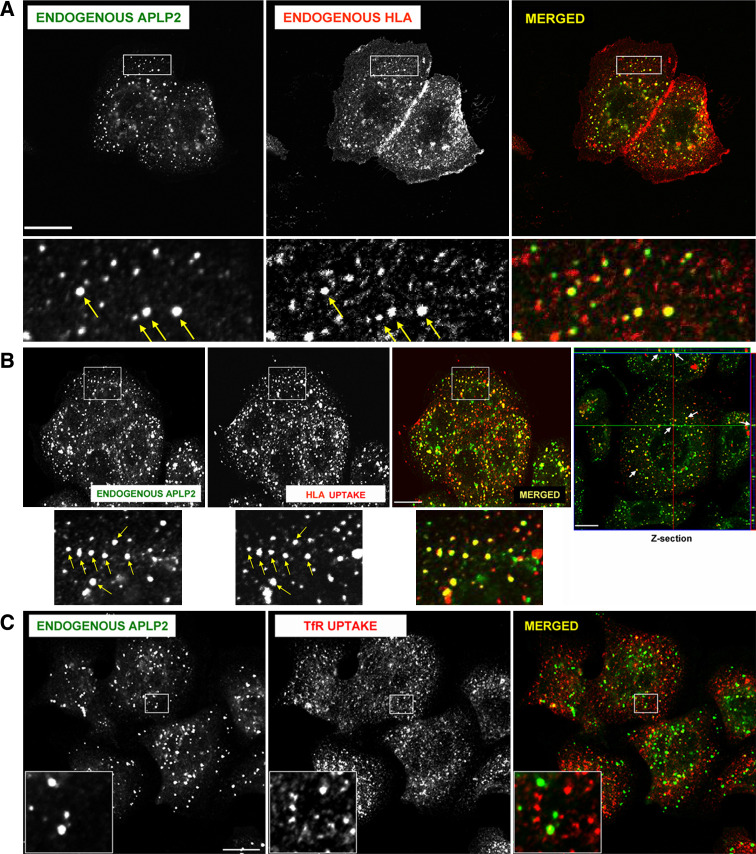
Fig. 2.
APLP2 molecules were co-localized with folded HLA class I molecules in vesicular structures in human pancreatic tumor S2-013 cells. a S2-013 cells were fixed with 4% paraformaldehyde in PBS for 10 min prior to incubation in staining solution (0.2% saponin, 0.5% bovine serum albumin in PBS) with mouse anti-HLA-A,B,C antibody (W6/32) and rabbit anti-APLP2 serum for 1 h at room temperature. The cells were then washed three times with PBS (5 min/wash) and incubated with fluorescently labeled secondary antibodies (Alexa Fluor 568 goat anti-mouse and Alexa Fluor 488 goat anti-rabbit antibodies) for 30 min at room temperature. Green APLP2, red folded HLA class I molecules, yellow co-localized APLP2 and HLA class I molecules. Yellow arrows indicate vesicles containing co-localized endogenous HLA class I and APLP2 molecules. More highly magnified images of areas shown in the larger boxes are shown below the boxes. b S2-013 cells were incubated with mouse anti-HLA-A,B,C antibody (W6/32) at 4°C, then warmed to 37°C for 30 min. The cells were treated with stripping solution (0.5% acetic acid, 500 mM NaCl) for 90 s to remove non-internalized surface-bound antibody, and fixed with 4% paraformaldehyde in PBS for 10 min. The cells were then incubated in staining solution with rabbit anti-APLP2 serum for 1 h at room temperature, washed three times (5 min/wash) with PBS, incubated for 30 min at room temperature with Alexa Fluor 488 goat anti-rabbit and Alexa Fluor 568 goat anti-mouse antibodies in staining solution, and washed three times with PBS (5 min/wash). More highly magnified images of areas shown in the larger boxes are shown below the boxes. Confirmation that APLP2 and endocytosed folded HLA class I molecules are located together in vesicles in S2-013 cells was obtained by taking z-section images. Serial z-section images were obtained every 0.4 μm, and the crosshairs and arrows depict common membrane structures on a representative photomicrograph containing both APLP2 (green) and human MHC class I (red). These data verify that the indicated APLP2- and HLA-containing structures are identical (yellow) and not merely overlaid. Arrows indicate vesicles with co-localized endogenous APLP2 and endogenous, endocytosed HLA class I molecules. c As a negative control, S2-013 cells were incubated with mouse anti-transferrin receptor antibody at 4°C, and then were warmed to 37°C for 15 min. The cells were treated with stripping solution (0.5% acetic acid, 500 mM NaCl) for 120 s to remove non-internalized surface-bound antibody, and fixed with 4% paraformaldehyde in PBS for 10 min. The cells were then incubated in staining solution with rabbit anti-APLP2 serum for 1 h at room temperature, washed three times (5 min/wash) with PBS, incubated for 30 min at room temperature with Alexa Fluor 488 goat anti-rabbit and Alexa Fluor 568 goat anti-mouse antibodies in staining solution, and washed three times with PBS (5 min/wash). The insets depict more highly magnified images of areas shown in the larger boxes. Green APLP2; red transferrin receptor. For (a), (b), and (c), images were analyzed on a Zeiss LSM 5 Pascal confocal microscope and bar 10 μm (Color figure online)

