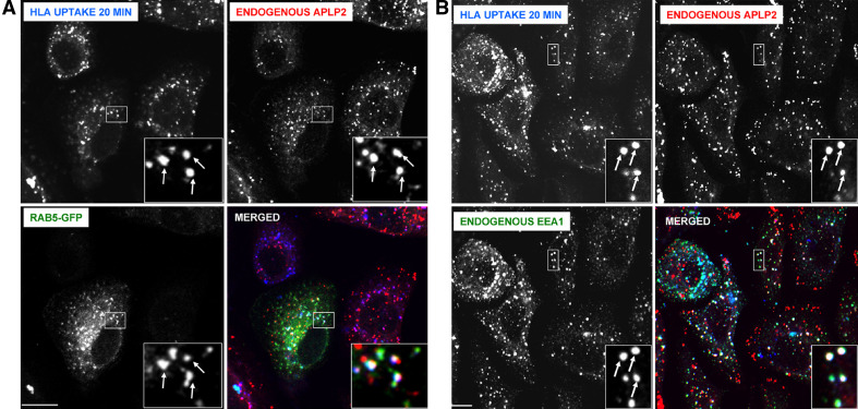
Fig. 3.
In pancreatic tumor S2-013 cells, APLP2 molecules were demonstrated to be co-localized with folded HLA class I molecules in vesicular structures containing markers of early endosomes. a S2-013 cells were transfected with Rab5-GFP using Effectene reagent. At 24 h post-transfection, the cells were incubated with mouse anti-HLA-A,B,C antibody (W6/32) at 4°C, then warmed to 37°C for 20 min. The cells were treated with stripping solution (0.5% acetic acid, 500 mM NaCl) for 120 s to remove non-internalized surface-bound antibody, and fixed with 4% paraformaldehyde in PBS for 10 min. The cells were then incubated in staining solution with rabbit anti-APLP2 serum for 1 h at room temperature, washed three times (5 min/wash) with PBS, incubated for 30 min at room temperature with Alexa Fluor 568 goat anti-rabbit and Alexa Fluor 405 goat anti-mouse antibodies in staining solution, and washed three times with PBS (5 min/wash). Blue HLA class I, red APLP2; green Rab5-GFP; white merged. Arrows indicate some of the vesicles with co-localized HLA class I, APLP2, and Rab5-GFP. b S2-013 cells were incubated with mouse anti-HLA-A,B,C antibody (W6/32, which is an IgG2a antibody) at 4°C, then warmed to 37°C for 20 min. The cells were treated with stripping solution (0.5% acetic acid, 500 mM NaCl) for 120 s to remove non-internalized antibody, and fixed with 4% paraformaldehyde in PBS for 10 min. The cells were then incubated for 30 min in staining solution with Alexa Fluor 405 goat anti-mouse antibody. After three washes of 5 min each with PBS, the cells were incubated for 1 h at room temperature in staining solution with rabbit anti-APLP2 serum and mouse anti-EEA1 antibody (IgG1). After an additional three washes in PBS (5 min/wash), the cells were incubated for 30 min at room temperature with Alexa Fluor 568 goat anti-rabbit antibody and Alexa Fluor 488 goat anti-mouse IgG1-specific antibody in staining solution, and washed three times with PBS (5 min/wash). Blue HLA class I; red APLP2; green EEA1; white merged. Arrows indicate some of the vesicles with co-localized HLA class I, APLP2, and EEA1. For both (a) and (b), images were analyzed on a Zeiss LSM 5 Pascal confocal microscope, bar 10 μm, and insets depict more highly magnified images of the areas shown in the larger boxes (Color figure online)

