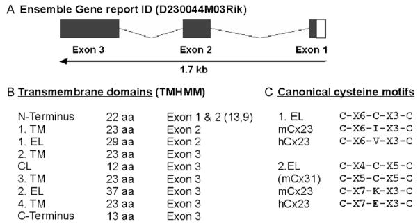
Fig. 1.
Sequence analyses of mouse Cx23. (A) Schematic drawing of the mouse Cx23 gene structure (1.7 kb) as deduced from the Ensemble Gene Report. The coding region of 618 nucleotides is distributed over three exons. (B) Size and exon-specific distribution of nine different putative domains of mouse Cx23 corresponding to transmembrane topology predicted by the HUSAR-derived subprogram TMHMM. (C) Comparison of the canonical cysteine motifs known from connexin genes (1.EL and 2.EL) with the cysteine motifs of mouse and human Cx23. The central cysteine in both presumably extracellular loops of Cx23 is missing. The distribution of the other conserved amino acid residues between the two flanking cysteines in the second extracellular loop is changed. C, cysteine, K, lysine, E, glutamate X, miscellaneous amino acid. EL, extracellular loop; CL, cytoplasmic loop; TM, transmembrane region.

