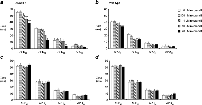
Fig. 4.
APD at 30%, 50%, 70%, and 90% repolarization quantified from MAP recordings made from the epicardium of KCNE1−/− (a) and WT (b) hearts during 8-Hz extrinsic pacing demonstrated a concentration-dependent reduction in APD90 and APD70 with nicorandil. In contrast, similarly obtained MAP recordings from the endocardium of KCNE1−/− (c) and WT (d) hearts demonstrated conserved APD values during perfusion with nicorandil (*P < 0.05; **P < 0.01; ***P < 0.001)

