
Figure 4. Differential activation of CaMKII in spines and dendrites by postsynaptic depolarization.
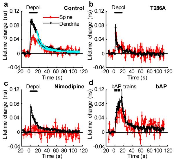
a, High temporal resolution measurements of the fluorescence lifetime change of Green-Camuiα in spines and dendrites in response to postsynaptic depolarization (0 mV, 16 s, indicated by the bar) in spines (red) and dendritic shafts (black). Average of 53 spines and 30 dendrites from 12 neurons. The cyan lines are a double exponential function obtained by fitting. The time constants are 11.3 s (84 %) and > 300 s (16%) for spines, and 6.8 s (60 %) and 43.7 s (40 %) for dendrites.
b, Fluorescence lifetime change of T286A mutant of Green-Camuiα. Average of 19 spines and 14 dendrites from 8 neurons.
c, Fluorescence lifetime change of Green-Camuiα in the presence of L-type VSCC inhibitor (20 μM Nimodipine). Average of 22 spines and 15 dendrites from 7 neurons.
d, Bursts of back-propagating action potentials (bAP) induced CaMKII activation at proximal dendrites (< 100 μm). We applied 8 bAP bursts, each 83 Hz for 0.5 s, at 2 s intervals. Average of 22 spines and 12 dendrites from 7 neurons).