
Figure 7.
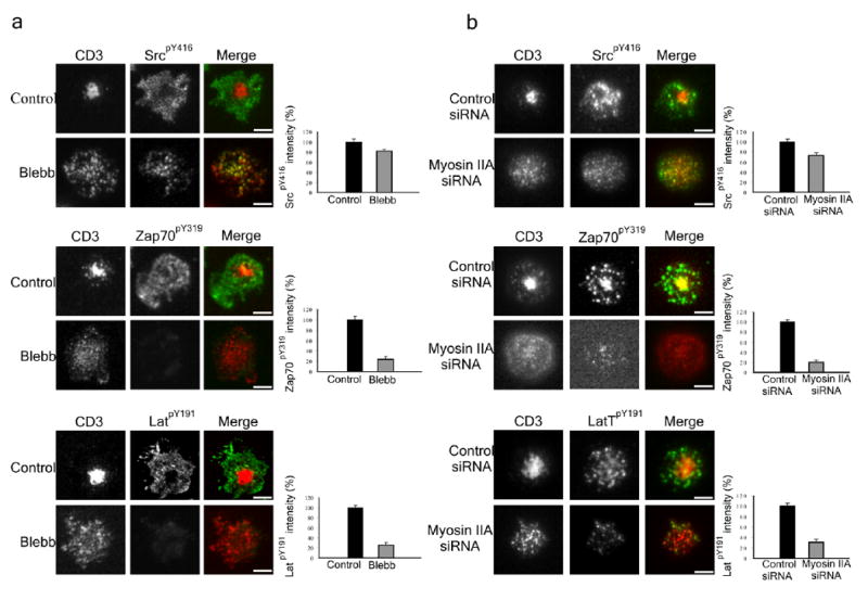
The effect of inhibiting or depleting myosin-II on signaling in T cells. (a) Control or blebbistatin pretreated Jurkat T cells were added to a planer lipid bilayer containing Alexa-568 labeled TCR antibody and ICAM1 for 25 min. Cells were then fixed and stained with antibodies against SrcpY416, ZAP70pY319 and LATpY191. Quantitative representation of relative protein phosphorylation is depicted on the right (n = 15 cells for each bar and error bars indicate standard deviation). (b) Primary human CD4+ cells treated with siRNA constructs either specific or non-specific for MYH9 gene were added to a planer lipid bilayer containing Alexa-568 labeled TCR antibody and ICAM-1 for 25 min. Cells were then fixed and stained with antibodies against SrcpY416, ZAP70pY319 and LATpY191. Myosin IIA depleted cells were determined by the lack of central TCR clustering as demonstrated in Fig. 6b. Quantitative representation of relative protein phosphorylation is depicted on the right (n = 15 cells for each bar).