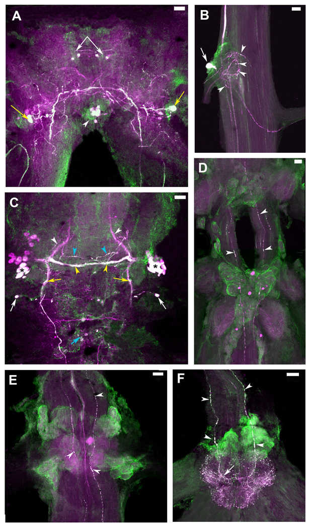
Figure 8. Colocalization of 5-HT1Mac and TH immunoreactivity in the CNS of the prawn.
5-HT1Mac immunoreactivity (ir) shown in green and Tyrosine Hydroxylase (TH) ir shown in magenta. TH ir is taken to represent dopamine (DA)-containing cells (see Materials & Methods). Cells and fibers showing immunoreactivity to both 5-HT1Mac and TH appear white. A: View of the brain, showing colocalization of both 5-HT1Mac and TH/DA in a group of four small-sized cells in the protocerebrum (white arrows), a pair of large- to medium-sized cells in the deutocerebrum (yellow arrows), and a cluster of 4–8 medium-sized cells in the tritocerebrum (white arrowheads). B: View of the circumesophageal ganglion (CEG), showing colocalization for both 5-HT1Mac and TH/DA in a large-sized cell (white arrow) that sends its axon towards the brain, as well as in terminal arborizations (white arrowheads) within the neuropil of the ganglion. C: View of the subesophageal ganglion (SEG), showing 5-HT1Mac and TH/DA colocalization in some (*), but not all, of the medium-sized cells within the bilateral clusters of the ganglion, in the fascicles of axons going to the contralateral (yellow arrowheads) side of the ganglion (but not in those going towards the CEG; white arrowheads), within longitudinal fibers communicating the SEG with the lower ganglia (yellow arrows), in some, but not all, of the horizontally-oriented fibers forming a ladder-like pattern (blue arrowheads), and in the small-sized cells on the edges of the ganglion (white arrows), inferior to the bilateral clustered cells, as well as in the centrally located cells (blue arrow) between the SEG and the first thoracic ganglion (T1). D: View of T3-T4, showing 5-HT1Mac and TH/DA colocalization only in the longitudinal fibers that traverse the ganglia (white arrowheads). E: View of the first abdominal ganglion (A1), showing 5-HT1Mac and TH/DA colocalization in the longitudinal fibers that traverse the ganglia (white arrowheads) and rather sparsely on the terminal arborizations within the neuropil. F: View of the sixth abdominal ganglion (A6), showing 5-HT1Mac and TH/DA colocalization in the expansive terminal arborization that forms at the end of the longitudinal fibers entering through the connective. All images shown were obtained from the ventral nerve cord of a male blue clawed prawn. All images are composites of optical slices of sets of confocal stacks spanning the full dorsal-ventral axis of the ganglia. The fluorescence has been digitally brightened, and several pieces of surface debris have been digitally removed. (Scale bars = 100 µm).

