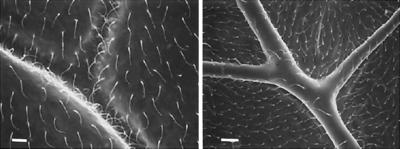
Figure 5.
Scanning electron microscope images of the ventral surface of the forewing of (Left) Taeniopteryx burksi (Taeniopterygidae; bar = 10 μm) and (Right) Paragnetina media (Perlidae; bar = 50 μm). Exposure to a vacuum causes the collapse of the vein cuticle over the entire ventral surface of T. burksi wings; no such collapse occurs in P. media, which has thicker ventral vein cuticles. There is no vein collapse on the dorsal surface of wings from either species (image not shown).

