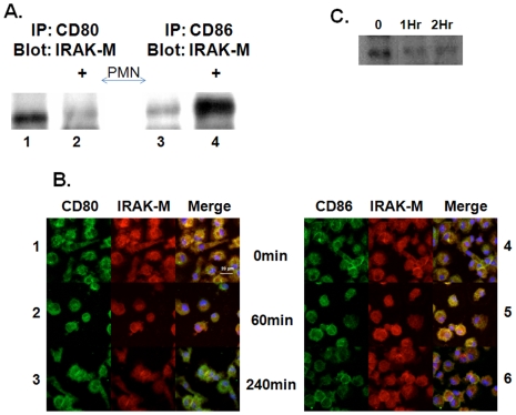
Figure 4. PMN membranes disassociate IRAK-M from CD80.
Macrophages were treated with PMN lipid rafts for various times Panel A. Co-IP demonstrates CD80 binds IRAK-M in resting PMA differentiated THP-1 macrophages, lane 1. One hour after lipid raft treatment IRAK-M disassociates from CD80, lane 2. CD86 binds little IRAK-M in resting macrophages, lane3. There is association of IRAK-M with CD86 one hour after lipid raft addition, lane4. Panel B. Confocal microscopy of resting monocyte derived macrophages at baseline (Row 1,4) or stimulated with PMN lipid rafts for 1 hr (Row 2,5) or 4 hrs (Row 3,6). IRAK-M (Red), CD80 (Left Panel-green) or CD86 (Right Panel-green) and co-localization (Yellow). Nucelar stain with DAPI (blue). Panel C. PMA differentiated THP-1 macrophages were stimulated with PMN lipids rafts and harvested at the designated time point. Whole cell extracts were IP for TRAF-6 and blotted for IRAK-M. All lanes (panel A and C) were normalized for protein prior to immunoprecipitation.

