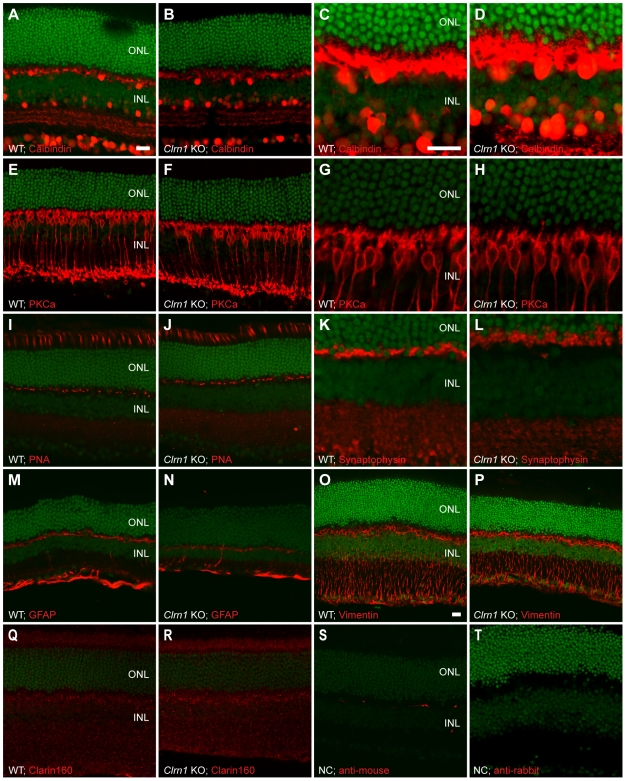
Figure 7. Immunohistochemical analysis of littermate WT (+/+) and Clrn1 KO (−/−) mouse retinas.
(A–D) WT and KO retinas stained with an antibody to Calbindin, shown at low (A and B) and high (C and D) magnifications. (E–H) WT and KO retinas stained with an anti-PKC-alpha antibody at low (E–F) and high (G–H) magnifications. (I,J) WT and KO retinas labeled with the rhodamine-conjugated peanut agglutinin (PNA) lectin. (K,L) WT and KO retinal sections immunostained with anti-Synaptophysin antibodies, shown at high magnification. (M–P) WT and KO retinal sections stained with intermediate filament antibodies to GFAP (M,N) and vimentin (O,P). (Q,R) Immunostaining with a candidate antibody made to CLRN1 (the Clrn1 gene product) in WT (Q) and KO (R) retinas. (S,T) Control images showing labeling by the secondary antibodies alone. Scale bar = 20 µm in all images. Scale bars: In A applies to A–B, E–F, I–J, M–N, and Q–T; in C applies to C–D, G–H, and K–L; in O, applies to O–P. INL = inner nuclear layer, ONL = outer nuclear layer. SYTOX Green was used as the nuclear counterstain.

