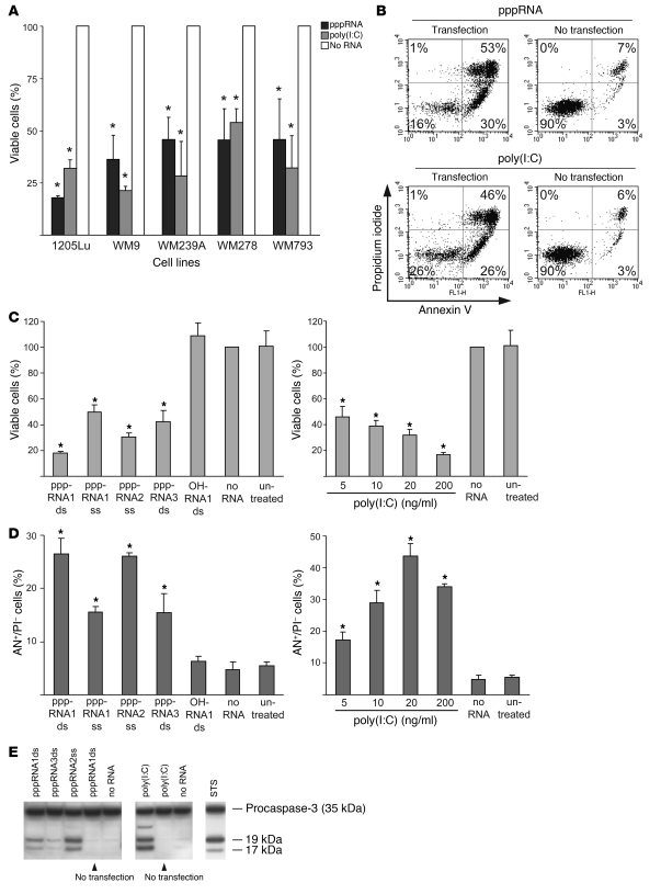
Figure 1. pppRNA and poly(I:C) induce apoptosis in melanoma cells.
(A) The viability of different melanoma cell lines was determined after transfection of in vitro–transcribed RNA (pppRNA) or poly(I:C) (20 ng/ml). Cells were analyzed 17 (pppRNA) or 24 hours [poly(I:C)] after transfection. Viability of cells treated with transfection reagent alone (mock-transfected cells; No RNA) was set to 100%. *P ≤ 0.05 compared with the same cell line treated with transfection reagent alone. (B) Fluorescence-activated cell sorting (FACS) analysis of apoptotic 1205Lu cells treated with pppRNA or poly(I:C) (200 ng/ml) for 24 hours either complexed with a liposomal transfection reagent (Transfection) or not (No transfection). Percentages indicate the portion of cells in each quadrant that defines annexin V– or propidium iodide–positive or –negative cells. Results are representative of 3 independent experiments. (C) Left: Viability of the metastatic melanoma cell line 1205Lu was measured 24 hours after transfection of different short in vitro–transcribed RNAs (pppRNA1–3) in double-stranded (ds) or single-stranded (ss) form or synthetic unconjugated RNAs (OH) with the same sequence. Sequences of pppRNAs are shown in Methods. Right: Viability of 1205Lu cells transfected with different doses of poly(I:C) (20–200 ng/ml) for 24 hours. Viability of mock-transfected cells was set to 100%. (D) FACS analysis of apoptotic 1205Lu cells treated with pppRNA or poly(I:C) as described in C. Annexin V–positive and propidium iodide–negative cells (AN+/PI–) are depicted. In A, C, and D, the mean ± SD of 3 independent experiments is shown. *P ≤ 0.05 compared with mock-transfected cells in C and D. (E) pppRNAs (pppRNA1–3) or poly(I:C) (5 ng/ml) were used with or without transfection reagent, and 1205Lu cells were analyzed 12 (pppRNA) or 24 hours [poly(I:C)] after treatment. Activation of caspase-3 was assessed by immunoblotting with an antibody specific for procaspase-3 (35 kDa) and its active cleaved subunits (17, 19 kDa). WM9 melanoma cells treated with 1 μM staurosporine (STS) for 5 hours served as a positive control for caspase cleavage. One representative of 3 independent experiments is shown.

