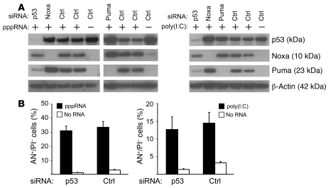
Figure 6. Role of p53 in apoptosis induced by RIG-I and MDA-5.
(A) 1205Lu cells were treated with pppRNA or poly(I:C) 48 hours after transfection of siRNAs specific for p53, Noxa, Puma, or control siRNA and analyzed by immunoblotting. Left: Treatment with pppRNA. Blots are representative of 3 independent experiments. Right: Treatment with poly(I:C) (20 ng/ml). Blots are representative of 2 independent experiments. β-Actin served as loading control. (B) 1205Lu cells were transfected with a p53-specific siRNA or control siRNA for 48 hours, treated with pppRNA (left) or poly(I:C) (5 ng/ml; right) or transfection reagent alone, and analyzed for apoptotic cells (AN+/PI–). Mean ± SD of 3 independent experiments is shown.

