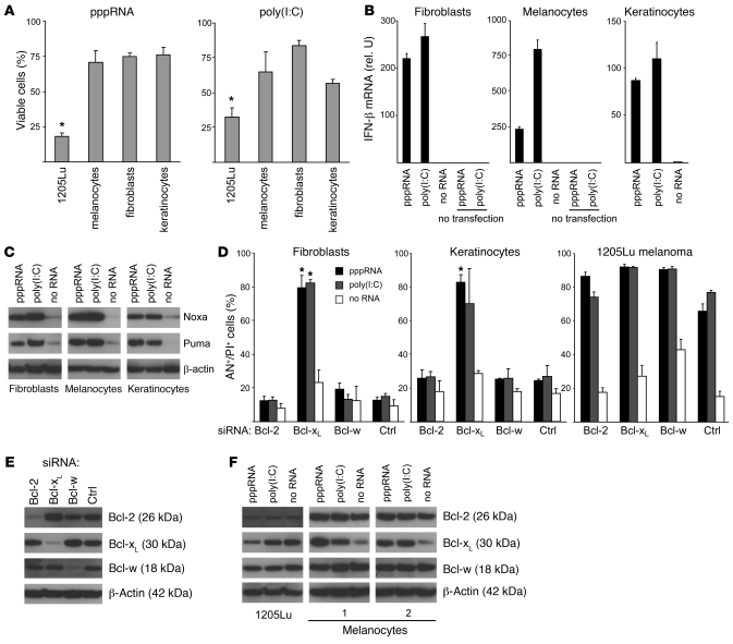
Figure 7. Increased apoptotic sensitivity of melanoma cells to RNA ligands of RIG-I and MDA-5.
(A) Cell viability of 1205Lu melanoma cells was compared with that of human melanocytes, primary human fibroblasts, or primary human keratinocytes. Cells were transfected with pppRNA or poly(I:C) (20 ng/ml) for 24 hours. The viability of mock-transfected cells was set to 100% for each cell type. Mean of 3 transfections of 1205Lu is indicated; the mean ± SEM of 2 or 3 donors measured in triplicate for primary cells is shown. *P ≤ 0.05 compared with melanocytes, fibroblasts, or keratinocytes. (B) Primary cells were treated with pppRNA, poly(I:C) (3 ng/ml) with or without transfection reagent, or with transfection reagent alone. IFN-β expression was analyzed by quantitative RT-PCR 17 hours after transfection. Mean ± SD of triplicate measurements of RNAs pooled from 3 (melanocytes) or 2 donors (fibroblasts and keratinocytes) is shown. (C) Primary cells were transfected with pppRNA for 17 hours or poly(I:C) (10 ng/ml) for 24 hours, and expression of Puma and Noxa protein was quantified by immunoblotting. Blots are representative of 3 independent experiments. (D) Primary fibroblasts, primary keratinocytes, or 1205Lu melanoma cells were treated with pppRNA, poly(I:C) (3 ng/ml), or transfection reagent alone 48 hours after transfection of siRNAs specific for antiapoptotic Bcl-2, Bcl-xL, Bcl-w, or control siRNA. Cell death was determined by FACS 17 hours after treatment with pppRNA or poly(I:C). Annexin V– and propidium iodide–positive cells are represented. Mean ± SD of 3 experiments with different donors for primary cells or different passages of 1205Lu cells is shown. *P ≤ 0.05, primary cells compared with cells transfected with control siRNA and the respective stimulus, pppRNA or poly(I:C). (E) Primary fibroblasts were treated with the indicated siRNAs and analyzed 48 hours after transfection by immunoblotting. Blots are representative of 3 independent experiments. (F) Expression of Bcl-2, Bcl-xL, and Bcl-w upon transfection with RIG-I and MDA-5 ligands. Melanocytes of 2 donors or 1205Lu melanoma cells were treated with pppRNA for 17 hours or poly(I:C) (10 ng/ml) for 24 hours. Expression was quantified by immunoblotting. Blots are representative of 3 independent experiments for 1205Lu melanoma cells. In C, E, and F, β-actin served as loading control.

