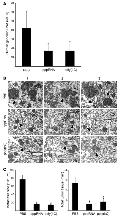
Figure 8. Therapeutic efficacy of RIG-I and MDA-5 ligands in immunodeficient mice.
(A) Groups of 3 NOD/SCID mice were challenged with 4 × 10⁵ 1205Lu melanoma cells and treated intravenously on days 3, 6, and 9 with pppRNA, poly(I:C), or PBS complexed with jetPEI as described in Methods. Human genomic DNA, representative for lung metastases, was measured in triplicate by quantitative PCR in lungs isolated at day 10. The relative amount of genomic DNA was expressed as a ratio of the amount of murine genomic DNA determined in the same lung sample. Mean ± SEM of each group is depicted. (B) Groups of 3 NOD/SCID mice were challenged with 4 × 10⁵ 1205Lu melanoma cells and treated intravenously on days 3, 6, 9, and 20 with pppRNA, poly(I:C), or PBS as described in Methods. Analysis was done on day 24. Representative lung sections after H&E staining of individual mice of each group are shown. 1205Lu metastases are indicated by black arrowheads. Scale bar: 100 μm. (C) Left: Metastasis size was calculated from the diameter in histological sections as described in Methods. Mean metastasis size was determined for each mouse, and the mean of each group ± SEM is shown. Right: Total tumor burden was calculated as the sum of all metastasis areas in each mouse as described in Methods. Mean ± SD of each group is shown. *P ≤ 0.05 compared with PBS-treated mice.

