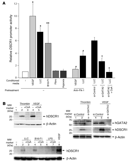
Figure 6. Modulatable DSCR1s promoter activity and protein expression in cultured microvascular endothelial cells.
(A) HMVECs were transiently transfected with DSCR-1–luc2cp. Cells were then transfected with siRNA or treated with anti–Flk-1 neutralizing antibody or CsA and subsequently exposed to conditioned medium. The results show the mean ± standard deviation of luciferase light units (relative to untreated cells) obtained in triplicate from 3 independent experiments. *P < 0.001, **P < 0.01 compared with control; †P < 0.001 compared with si-Control plus LLC-conditioned medium. #P < 0.001 compared with no-pretreatment controls. (B) Top left: HMVECs were pretreated with vehicle control (lanes 1, 2, and 4) or 1 μM CsA (lanes 3 and 5) and then incubated in the absence (lane 1) or presence of thrombin (lanes 2 and 3) or VEGF (lanes 4 and 5). Cell lysates were subjected to immunoblotting with DSCR-1 antibody. Top right: HMVECs were transfected with control siRNA (lanes 1, 2, 4, and 5) or GATA2 siRNA (lanes 3 and 6) and then treated without (lanes 1 and 4) or with thrombin (lanes 2 and 3) or VEGF (lanes 5 and 6). Cell lysates were subjected to immunoblotting with DSCR-1 and GATA2 antibodies. Bottom: HMVECs were pretreated with vehicle control (lanes 1, 2, 4, 6, and 8) or 1 μM CsA (lanes 3, 5, and 7) and then incubated in the absence (lane 1) or presence of conditioned medium from LLC (lanes 2 and 3), B16-F1 (lanes 4 and 5), LPS (lanes 6 and 7), or VEGF (lane 8). Membranes were stripped and reprobed with anti–β-actin antibody.

