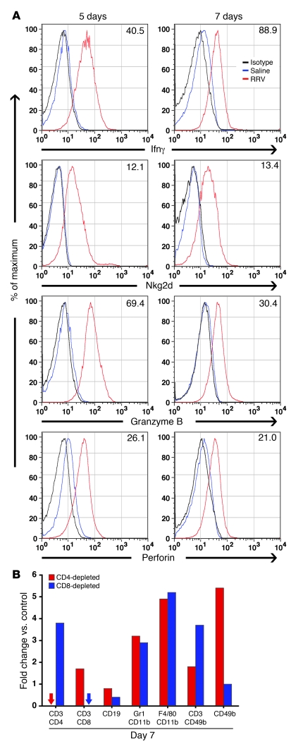
Figure 3. RRV-induced activation of hepatic NK cells after RRV challenge.
(A) Flow cytometry histograms showing the expression of Ifnγ, Nkg2d, granzyme B, and perforin by hepatic CD49b+ (NK) cells at 5 and 7 days after RRV or saline injection. For each individual cytokine/protease, the number represents the mean fluorescence intensity induced by RRV minus the median fluorescence intensity of the appropriate saline control. n = 3 livers per group per time point; % of maximum (vertical axis) represents a normalization of the number of events in specific quadrants of the flow cytometry grid. (B) Flow cytometric quantification of hepatic mononuclear cells is represented as fold changes in RRV-inoculated relative to saline-control mice that had been depleted of CD4+ or CD8+ cells (cells isolated at 7 days after injection of saline or RRV). n = 3–4 for each group; arrows indicate nondetectable cells; the horizontal axis shows the surface markers identifying specific cell types.

