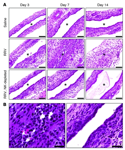
Figure 7. Prevention of bile duct injury and obstruction by NK cell depletion.
H&E staining of longitudinal sections of murine extrahepatic bile ducts at different time points after saline or RRV injection in the first day of life; the “RRV, NK depleted” group of mice was also injected daily with NK cell–depleting serum (A). (B) The photomicrograph on the left is a higher-magnification image of the middle panel in A showing the inflammatory obstruction of the duct lumen 7 days after RRV, while the photograph on the right shows the intact epithelium and unobstructed duct lumen 7 days after RRV infection of an NK cell–depleted mouse. Asterisks indicate the bile duct lumen. Scale bars: 50 μm.

