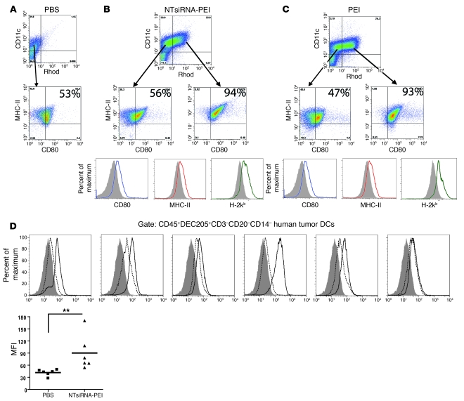
Figure 2. Engulfment of PEI-based nanocomplexes induces maturation of tumor-associated DCs in vivo and in situ.
(A) Expression of CD80 and MHC-II on tumor-associated DCs from mice bearing ID8-Defb29/Vegf-A tumors for 3 weeks and injected with PBS. (B) Rhodamine-labeled NTsiRNA-PEI or (C) equivalent amounts of rhodamine-labeled PEI alone (see Methods) were intraperitoneally injected into mice bearing ID8-Defb29/Vegf-A tumors for 3 weeks. Expression of CD80, MHC-I, and MHC-II on tumor DCs that engulfed the nanoparticles or PEI was analyzed by FACS 3 days later. Rhod, rhodamine-labeled PEI. Filled histograms represent expression on DCs that did not engulf nanoparticles or PEI. Open histograms indicate expression by DCs that engulfed nanoparticles or PEI in the same host. Data are representative of 3 mice per group in 4 independent experiments. In A–C, the percentage of tumor-associated DCs coexpressing MHC-II and CD80 is indicated. (D) siRNA-PEI nanoparticles induced maturation of human tumor DCs. Leukocytes from 6 unselected dissociated stage III human ovarian tumors were enriched by Ficoll, 5 × 10⁶ cells were incubated in 5 ml RPMI containing 100 μl PBS (dotted histograms) or 100 μl NTsiRNA-PEI (open histograms; see Methods), and the levels of CD80 on CD45+DEC205+CD3–CD20–CD14– tumor DCs were analyzed by FACS 18 hours later. Filled histograms represent staining with isotype control antibodies. MFI, mean fluorescence intensity of CD80 staining. Data points and horizontal bars denote individual values and means, respectively. **P < 0.01 (Mann-Whitney).

