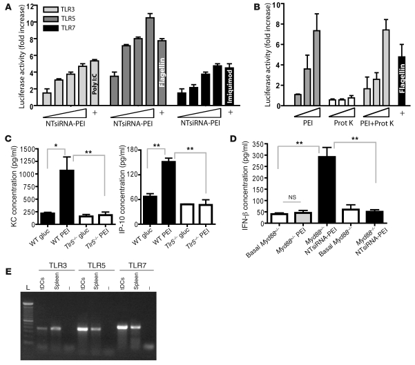
Figure 4. siRNA-PEI nanoparticles stimulate multiple TLRs in vitro and in vivo.
(A) siRNA-PEI nanocomplexes dose-dependently stimulated TLR3, TLR5, and TLR7 in vitro. Cotransfected HEK293 cells were stimulated with increasing amounts of siRNA-PEI nanoparticles or positive control agonists (+) as described in Methods. Data are representative of 4 independent experiments. (B) PEI was sufficient to stimulate TLR5 in vitro in a dose-dependent manner. HEK293 cells expressing TLR5 and harboring an NF-κB–dependent luciferase reporter plasmid were stimulated with increasing amounts of PEI, proteinase K (Prot K), or PEI treated overnight at 55°C with proteinase K (1 mg/ml), and luciferase activity was measured as described in Methods. Data are representative of 3 independent experiments. (C) PEI induced rapid cytokine secretion in vivo in a TLR5-dependent manner. Naive WT or Tlr5–/– mice were intraperitoneally injected with 5% glucose (gluc) or linear PEI, and serum levels of KC and IP-10 were determined 2 hours later by cytokine assay (see Methods). Data are representative of 4 mice per group, 2 independent experiments. (D) PEI-complexed siRNA induced MyD88-dependent secretion of IFN-β at tumor locations. Ascites from Myd88+/– or Myd88–/– mice bearing advanced ID8-Defb29/Vegf-A ovarian tumors were collected prior to (Basal) and 3 hours after intraperitoneal administration of PEI or NTsiRNA-PEI, and levels of IFN-β were analyzed by ELISA. Data are representative of 4 mice per group, 2 independent experiments. (E) CD45+CD11c+MHC-II+ tumor DCs were sorted from the peritoneal cavity of mice bearing ID8-Defb29/Vegf-A tumors, and TLR expression was confirmed by RT-PCR. Error bars in A–D denote SEM. *P < 0.05; **P < 0.01.

