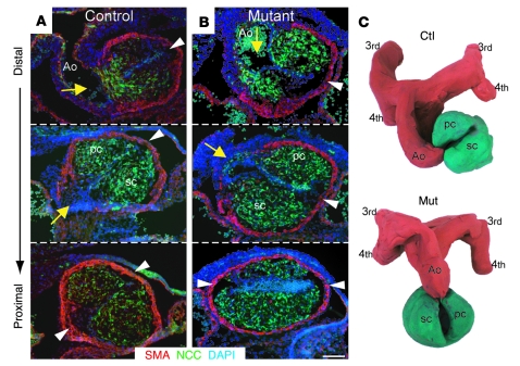
Figure 4. Impaired outflow tract rotation in conditional Fak mutant embryos.
Distal-to-proximal frontal sections through the conotruncus of E11 control (A) and mutant (B) embryos, at the level of aortic branching from the outflow tract. Note abnormal positioning of the aorta (yellow arrows) in mutants, which is in a more dextroposed position as compared with control littermates. Arrowheads point to conotruncal cushion limits. Red shows SMA, green (GFP) shows NCCs migrating into the outflow tract, and blue stains show nuclei (DAPI). (C) Clay models of E11 outflow tracts based on frontal sections of Z/EG mutants and control littermates, showing the abnormal rotation of the outflow tract in mutant embryos. Cryostat frontal sections used for the construction of these models were 30 μm apart and triple stained with GFP (for NCC), SMA, and DAPI. Representative sections are illustrated in Supplemental Figure 5. pc, parietal cushion; sc, septal cushion; 3rd, third aortic arch. Scale bar: 100 μm.

