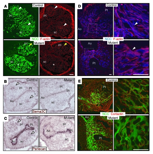
Figure 5. Abnormal NCC organization and morphology in conditional Fak mutant embryos.
(A–C) Transverse outflow tract sections of control and mutant E11.0 embryos. (A) Compared with control, GFP (green, NCCs) and phalloidin staining (red) demonstrate an abnormal organization of NCCs in conotruncal cushions of mutants. Note highly condensed mesenchyme of NCCs in the center of conotruncal cushions in control littermates (white arrowheads). In mutants, in contrast, these condensed structures are not found (asterisk) or they are disorganized and mislocalized to the edges of conotruncal cushions, near the myocardial layer (yellow arrowhead). (B and C) Semaphorin 3C (Sema3C) and perlecan in situ hybridization show reduced expression in the conotruncal cushions of Fak-deficient E11 outflow tracts (arrowheads). (D and E) Cryostat sections of E11.0 embryos at the level of the aorticopulmonary septum. (D) Compared with control, Fak-deficient NCCs (blue) show a rounder morphology and a disorganized actin cytoskeleton, demonstrated by phalloidin staining (red). Right panels show higher magnification images from boxed areas in left panels. Note that in control NCCs, F-actin is organized in parallel fibers as opposed to Fak-deficient NCCs (arrowheads). (E) Fak-deficient NCCs (green) show reduced cortactin (red) localization to the cell periphery in the aorticopulmonary septum compared with control littermates. Right panels show higher magnification images from boxed areas in left panels. Scale bars: 50 μm (A–C); 30 μm (D and E).

