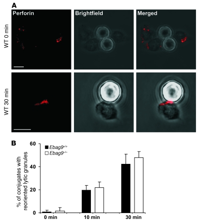
Figure 13. EBAG9 deletion does not affect formation of the immunological synapse.
(A) CTLs obtained from MLR (day 6–7) were mixed with the anti-CD3–coated latex beads and incubated for 0, 10, or 30 minutes at 37°C. Conjugates were plated on coverslips, fixed, and permeabilized. Perforin was stained with a biotinylated anti-perforin antibody. Polarization of perforin toward the contact site of CTLs and microbeads was assessed by confocal microscopy and quantified by random selection of conjugates. Those conjugates showing a distinct perforin immunofluorescence at the T cell–bead contact site were considered polarized. Beads are visible in Brightfield images as opaque round structures. One representative image of WT CTLs is shown. Scale bars: 5 μm. (B) Bars show statistical analysis from 3 independent experiments with at least 100 cells evaluated. Data represent mean ± SD.

