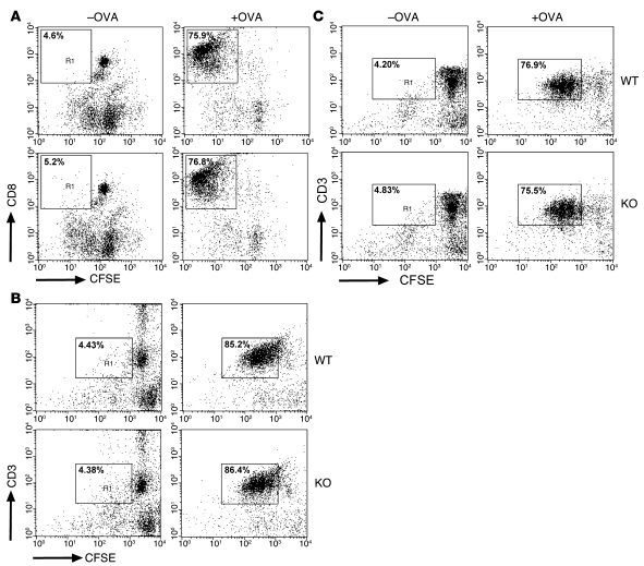
Figure 5. Similar efficiency of EBAG9-deficient and WT DCs for priming of OVA-specific T cells in vitro.
(A) DCs derived from Ebag9+/+ and Ebag9–/– mice (H-2b haplotype) were pulsed overnight with 100 μg/ml OVA protein. CFSE-labeled naive OT-I T cells (1 × 10⁵) were stimulated with pulsed or unpulsed DCs (1 × 10⁴) for 3 days. Cells were analyzed by flow cytometry, and T cells were gated for the expression of the clonotypic TCR or CD8a and CFSE. Shown is a representative FACS plot of gated CD8+ cells. Proliferation of T cells was evaluated according to CFSE dilution. Numbers indicate the percentage of proliferated CD8+ T cells within the gate. Representative data from 2 independent experiments with DCs from at least 6 animals per group. No significant differences were seen. (B) DCs (H-2d haplotype) were generated and pulsed with OVA protein (75 μg/ml) as described in A, followed by a 3-day coculture with CFSE-labeled OT-II CD4+ T cells. T cells were gated as described in A. Numbers indicate the percentage of proliferated CD3+ T cells within the gate. Shown are representative data from 3 independent experiments. DCs were taken from at least 6 animals per group. (C) DCs (H-2d haplotype) were pulsed for 2 hours with OVA-derived peptide (5 μg/ml), followed by a coculture with DO11.10 CD4+ T cells. T cells were gated as described in A. Shown are representative data from 4 independent experiments. DCs were taken from at least 9 animals per group. Percentages indicate the percentage of proliferated CD3+ T cells within the gate.

