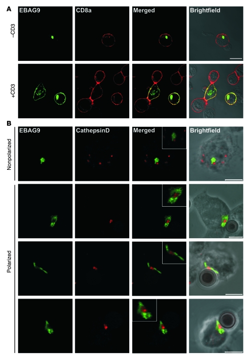
Figure 8. Activation-dependent intracellular redistribution of EBAG9 in CTLs.
(A) EBAG9 redistributes toward the plasma membrane upon T cell activation. For non-polarized activation via the TCR, EBAG9-GFP–transduced T cells were cultured on plate-bound anti-CD3ε and anti-CD28 mAbs (+CD3) or left untreated (–CD3). Cells were fixed and stained with an anti-CD8a antibody without permeabilization. Colocalization between EBAG9 and CD8a was quantified by calculating the Pearson’s correlation coefficient. At least 10 cells were analyzed in 2 independent experiments. Scale bar: 10 μm. (B) EBAG9 moves to the immunological synapse upon polarized stimulation of CTLs. CTLs were generated and transfected with EBAG9-GFP as described in Methods. CTLs were mixed with CD3/CD28-precoated Dynabeads, incubated for 5 minutes at 37°C, plated on coverslips, and incubated for another 30 minutes before fixation and permeabilization. Lytic granules were stained with anti–cathepsin D antibody (red). Representative images of non-conjugated and non-polarized T cells are shown in the top row. Polarized T cells with conjugated beads are shown in bottom 3 rows. Data are from 1 experiment with CTLs from 3 animals. At least 20 conjugates were analyzed (r = –0.10 ± 0.18). Scale bars: 5 μm. Insets have additional magnifications of 2- to 3-fold.

