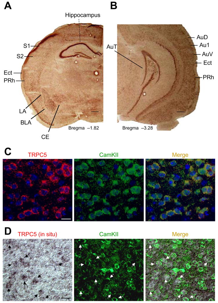
Figure 1. TRPC5 distribution in mouse brain.
(A, B) In situ hybridization of TRPC5-mRNA in the amygdala, hippocampus, somatosensory cortex, and auditory cortex. LA, lateral nucleus of the amygdala; BLA, basolateral nucleus of the amygdala; CE central nucleus of the amygdala; S1, primary somatosensory cortex; S2, secondary somatosensory cortex; AuD, secondary auditory cortex, dorsal; Au1, primary auditory cortex; AuV, secondary auditory cortex, ventral; Ect, ectorhinal cortex; PRh, perirhinal cortex. Scale bar, 1mm. (C), TRPC5 (left) and CaMKIIα (middle, a marker of pyramidal neurons) colocalize in the lateral nucleus of the amygdala. Scale bar, 25 μm. (D) TRPC5 (left, in situ hybridization) and anti-CaMKIIα (middle) co-localize in the majority of pyramidal cells of the auditory cortex. Arrows indicate cells expressing both TRPC5 and CaMKIIα. Scale bar, 50 μm.

