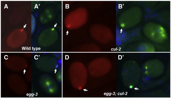
Fig. 3.
Meiotic cytoplasmic MEI-1 levels are not altered in cul-2 embryos. (A–D) Embryos stained with anti-MEI. (A′–D′) Anti-tubulin staining of the same embryos. In each panel, levels in meiotic embryos are compared to the mitotic embryo present in the same field. (A, A′) Wild-type cytoplasmic MEI-1 staining is higher in the meiotic embryo than a later-stage embryo. (B, B′) cul-2 has higher levels of cytoplasmic MEI-1 at meiosis than mitosis, and levels at both divisions are higher than corresponding wild-type embryos. (C, C′) egg-3 (RNAi) embryos show similar cytoplasmic MEI-1 levels at meiosis and mitosis, indicative of premature MEI-1 degradation. (D, D′) cul-2; egg-3(RNAi) embryos have similar levels of cytoplasmic MEI-1 at meiosis and mitosis, like egg-3 alone, but cytoplasmic levels at both divisions are increased, as seen with cul-2 single mutants. Note that in all panels meiotic spindles (arrows) are present and contain MEI-1.

