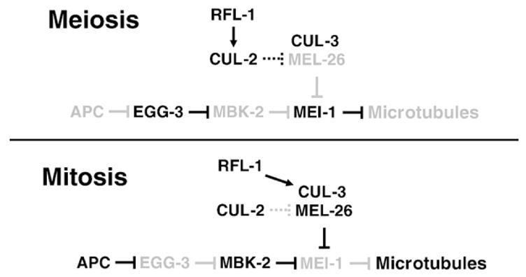
Fig. 5.

Model of MEI-1 regulation during the meiosis to mitosis transition. Low levels of protein or activity are indicated by lighter shading. During meiosis, CUL-2 (this report) and EGG-3 (Stitzel et al., 2007) keep MEL-26 and MBK-2 activities low, respectively, allowing MEI-1 to accumulate and sever microtubules. It is not known if the interaction between CUL-2 and MEL-26 is direct, and so this interaction is shown with stippled lines. During mitosis the situation is reversed and MEI-1 is degraded, allowing formation of longer microtubules. The kinase that acts in concert with the CUL-3/MEL-26 ubiquitin ligase and the ubiquitin ligase acting with MBK-2 are unknown.
