
Figure 4. A model for the organization of CEP55-ESCRT and CEP55-ALIX complexes in the midbody.
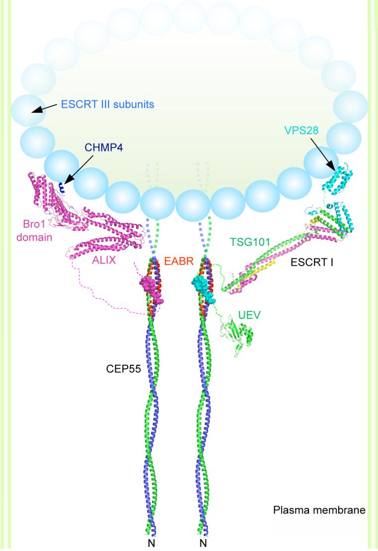
Model of the N-terminal half of the CEP55 structure docked to the ESCRT-I core (24) and UEV domain(21) and ALIX(25) structures as described in the on line methods supplement. The crystallized EABR portion of the CEP55 coiled coil is highlighted in red. The structure of the C-terminal domain of the yeast Vps28 subunit of ESCRT-I is shown (27) as a putative binding site for Vps20, the yeast ortholog of the human ESCRT-III subunit CHMP6. The binding site on the Bro1 domain of ALIX for the C-terminal helix (blue) of the CHMP4 subunit of ESCRT-III is shown (26, 28). A schematic of an ESCRT-III circular array (15) is shown. The width of the membrane neck is not to scale.