
Figure 1.

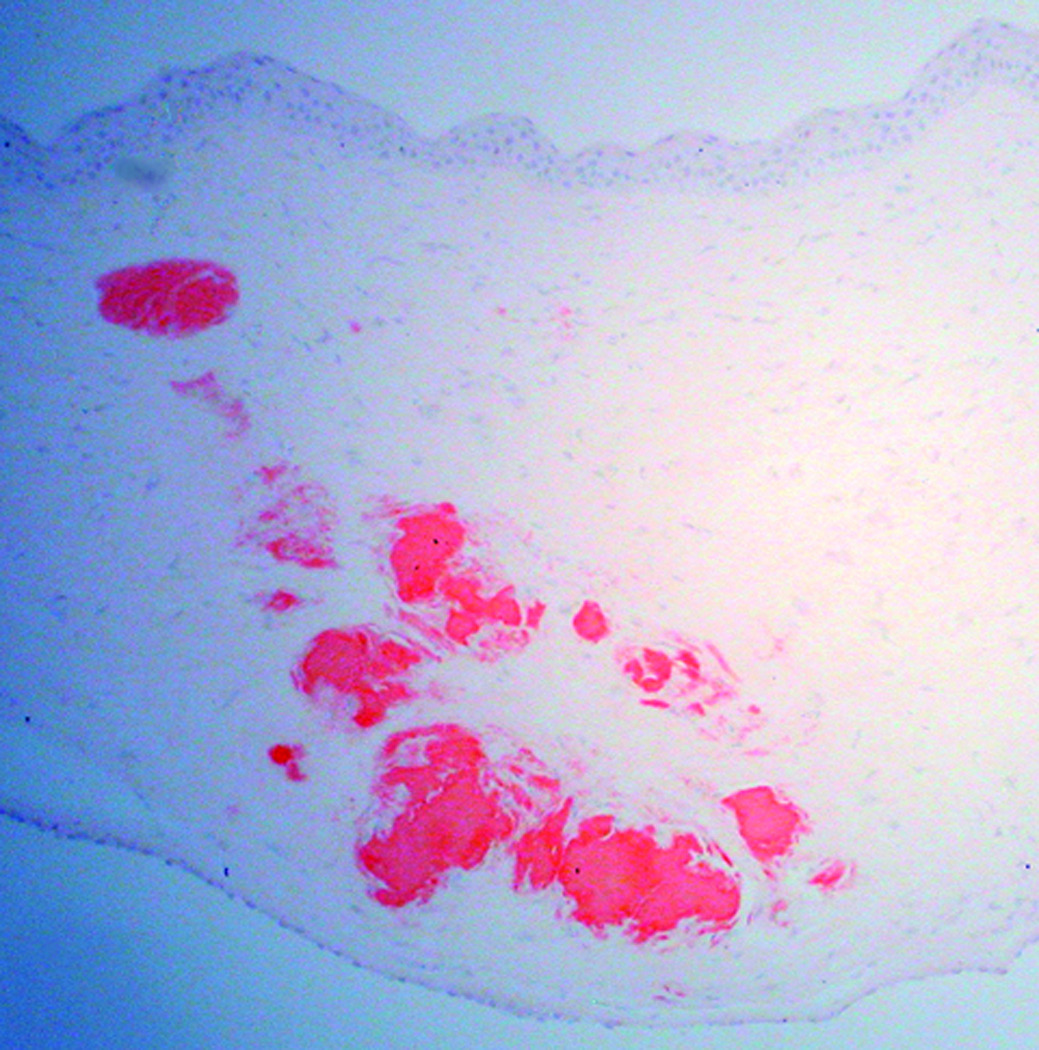
Congo red-stained sections of the right cornea from a 74-year-old man (Case 1 - Figure 1a) and the left cornea from a 37-year-old man (Case 2 - Figure 1b) who both underwent corneal transplantation for keratoconus. Red deposits that extend from the anterior to the deep stroma are noted in each section (original magnification, 200x).