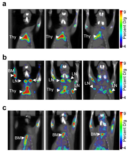
Figure 4. [18F]FAC microPET/CT allows visualization of increased lymphoid mass in systemic autoimmunity and can be used to monitor immunosuppressive therapeutic interventions.
Images are 60 minutes after i.v. injection of [18F]FAC and show three 1 mm thick coronal slices from (a) wild-type (C57BL/6J) and B6.MRL-Faslpr/J (b) before and (c) after treatment with DEX. [18F]FAC positive LNs were scored blindly. Thy, thymus; LN, lymph nodes; BM, bone-marrow. Results are representative of two independent experiments.

