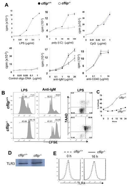
Figure 4. Proliferation and Ab responses in b-cflip−/− B cells.
(A). Splenic and lymph node B cells were stimulated with the indicated agonists. Proliferation was determined by the incorporation of radioactivity of [3H] thymidine (cpm, counts per minute). (B). B cell division kinetics were analyzed by CFSE dilution assays. At 48 h after stimulation, CFSE-labeled B cells were stained with 7AAD and analyzed by flow cytometry. The percentages of cell death are indicated by the 7AAD+ populations. (C) Cell death was determined at various times following LPS stimulation and death curves generated. (D) TLR3 expression in B cells was determined by western blotting. (E) TLR4 expression in B cells was analyzed by flow cytometry.

