
Figure 6.
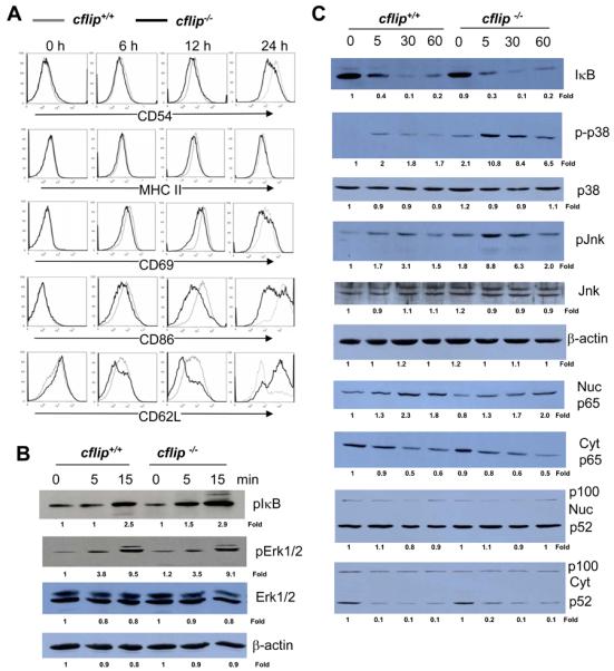
(A) Analysis of LPS-induced expression of activation markers and costimulatory proteins by flow cytometry. (B and C) B cells were stimulated with LPS, and western blotting was performed using Abs specific for pIκB, IκB, pErk1/2, Erk1/2, p65, p-p38, p38. pJnk, Jnk, and NF-κB p100/52. The values shown below each gel indicate folds of changes normalized against unstimulated sample (0 h) and loading controls (β-actin). Cyt: cytomplasmic; Nuc: nuclear.