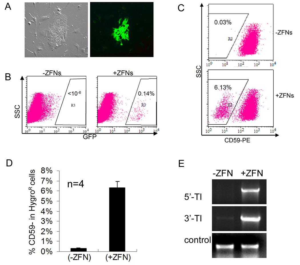
Figure 4. HR-mediated gene targeting of EGFP and PIG-A genes in female human MP2 iPS cells.
(A) Microscopy of corrected (GFP+) MP2 iPS cells harboring the EGIP* reporter, 5 days after Nucleofection with GFP-specific ZFNs.
(B) FACS analysis of EGFP gene correction in MP2 iPS cells with or without GFP-specific ZFNs. Dot plots were gated on TRA-1-60+ cells.
(C) PIG-A gene targeting in MP2 iPS cells with or without PIG-A specific ZFNs was performed as described in Figure 3A using the 1:5 ratio of Donor:ZFN DNA. After Nucleofection and hygromycin B selection, TRA-1-60+ (human iPS/ES) cells were analyzed for the presence or absence of CD59 (a GPI-AP). Representative FACS dot plots are shown.
(D) Summary of FACS analysis of CD59- MP2 iPS cell populations after PIG-A gene targeting from 4 independent experiments.
(E) PCR confirms the targeted integration into the PIG-A locus in MP2 iPS cells.

